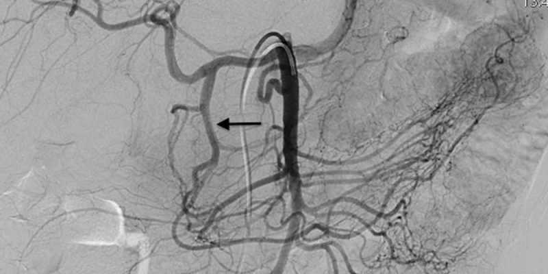
Renal arteriography is a special x-ray of the blood vessels of the kidneys.
How the Test is Performed
This test is done in the hospital. You will lie on an x-ray table.
Health care providers often use an artery near the groin for the test. Occasionally provider may use an artery in the wrist. Your provider will:
- Clean and shave the area.
- Apply a numbing medicine to the area.
- Place a needle into the artery.
- Pass a thin wire through the needle into the artery.
- Take out the needle.
- Insert a long, narrow, flexible tube called a catheter in its place.
The radiologist directs the catheter into correct position using x-ray images of the body. An instrument called fluoroscope sends the images to a TV monitor, which the provider can see.
The catheter is pushed ahead over the wire into the aorta (main blood vessel from the heart). It then enters into the kidney artery. The test uses a special dye (called contrast) to help the arteries show up on the x-ray. The blood vessels of the kidneys are not seen with ordinary x-rays. The dye flows through the catheter into the kidney artery.
X-ray images are taken as the dye moves through the blood vessels. Saline (sterile salt water) containing a blood thinner may also be sent through the catheter to keep blood in the area from clotting.
The catheter is removed after the x-rays are taken. A closure device is placed in the groin or pressure is applied to the area to stop the bleeding. The area is checked after 10 or 15 minutes and a bandage is applied. You may be asked to keep your leg straight for 4 to 6 hours after the procedure.

